Smooth Muscle Diagram - Muscle Tissue Types Learn Muscular Anatomy. You will have some basic understanding of the appearance referring to the below smooth muscle diagram. It is layered in a distinctive pattern of circular layers. Smooth muscles have a much stronger ability to contract than skeletal. Diagram of artery with smooth muscle identification understanding smooth muscles. It is the pen diagram of skeletal, smooth and cardiac muscle for class 10, 11 and 12.
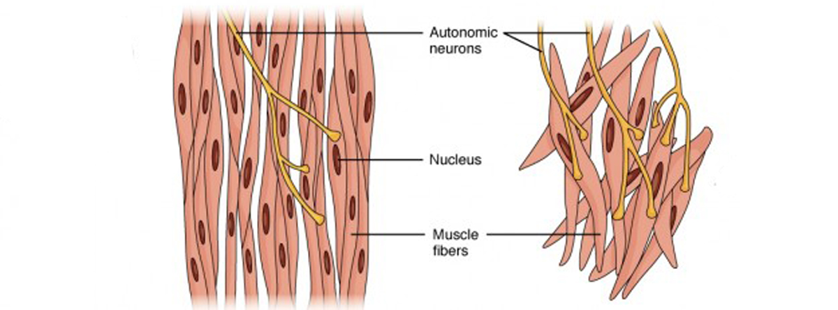
These cells have fibers of actin and myosin which run through the cell and are supported by a framework of other proteins. The cells stick together and are connected by specialised cell junctions, called gap junctions. 2 types of smooth muscle each fiber can contract independently and is usually innervated by a single nerve ending. This is just a diagram of how the human muscle looks under all the tissue and skin. Smooth muscle is made up of cells that contain a single central nucleus.
Smooth Muscle Definition And Examples Biology Online Dictionary from nitrocdn.com Diaphragm is also a skeletal muscle. Also, after activation of the receptors there is a long process in order to elicit an action potential, involving second. These cells have fibers of actin and myosin which run through the cell and are supported by a framework of other proteins. Diagram of smooth muscle contraction, smooth cardiac and skeletal muscle diagram, smooth muscle cell diagram, smooth muscle cell picture. Smooth muscle diagram labeled new epimysium learn schematic diagram. • smooth muscles respond to stretch only briefly, and then adapts to its new length • the new length however, retains its original _____ seconds or minutes after it has been elongated or shortened (e.g. It is layered in a distinctive pattern of circular layers. 2 types of smooth muscle each fiber can contract independently and is usually innervated by a single nerve ending.
Learn vocabulary, terms, and more with flashcards, games, and other study tools.
The cardiac muscle is only found in the heart wall. Muscle anatomy ankle 12 photos of the muscle anatomy ankle ankle anatomy muscle tendon, foot ankle. In addition, the contractile state of smooth muscle is controlled by hormones, autocrine/paracrine agents, and other local chemical signals. Smooth muscle makes up the walls of hollow organs, respiratory passageways, and blood vessels. Smooth muscle (also known as visceral muscle due to the locations in which they are present ) is one of the three main types of muscle tissue that exist in the human body. It is layered in a distinctive pattern of circular layers. Smooth muscle diagram, find out more about smooth muscle diagram. Smooth muscle is a type of tissue found in the walls of hollow organs, such as the intestines, uterus and stomach. • smooth muscles respond to stretch only briefly, and then adapts to its new length • the new length however, retains its original _____ seconds or minutes after it has been elongated or shortened (e.g. Smooth muscle contracts under certain stimuli as atp is freed. Smooth muscle diagram class 9. Vascular smooth muscle is the type of smooth muscle that makes up most of the walls of blood vessels. It constitutes much of the musculature of
Vascular smooth muscle refers to the particular type of smooth muscle found within, and composing the majority of the wall of blood vessels. This diagram shows a few of the cells that can be seen in the stained section below. It is the pen diagram of skeletal, smooth and cardiac muscle for class 10, 11 and 12. Beta 2 receptors are also on small coronary arterioles thus increasing hormonally induced blood flow within the musculature of the heart. It is the main muscle of respiration.
1 from The smooth muscles perform the functions in the contrast of other types of muscles. The cells are spindle shaped, and the nucleus is central. Smooth muscle tissue, unlike striated muscle, contracts slowly and automatically. In addition, the contractile state of smooth muscle is controlled by hormones, autocrine/paracrine agents, and other local chemical signals. Diagram of artery with smooth muscle identification understanding smooth muscles. It is layered in a distinctive pattern of circular layers. You will have some basic understanding of the appearance referring to the below smooth muscle diagram. The cardiac muscle is only found in the heart wall.
It is layered in a distinctive pattern of circular layers.
Learn vocabulary, terms, and more with flashcards, games, and other study tools. Smooth muscle diagram labeled new epimysium learn schematic diagram. Related posts of smooth muscle diagram labeled back muscle chart. Smooth muscle anatomy smooth muscle tissue is also known as visceral muscle tissue. Vascular smooth muscle is the type of smooth muscle that makes up most of the walls of blood vessels. The smooth muscle contraction is much slower than in the striated muscle primarily due to the presence of g protein coupled ligand receptors instead of ion channel coupled ligand gated receptors present in striated muscle. • smooth muscles respond to stretch only briefly, and then adapts to its new length • the new length however, retains its original _____ seconds or minutes after it has been elongated or shortened (e.g. Muscle anatomy ankle 12 photos of the muscle anatomy ankle ankle anatomy muscle tendon, foot ankle. Smooth muscle diagram, find out more about smooth muscle diagram. Vascular smooth muscle refers to the particular type of smooth muscle found within, and composing the majority of the wall of blood vessels. This smooth muscle can be found surrounding the walls of the blood vessels, the bronchioles in the lungs, and the sphincter muscles used in the gi tract.the gi tract, which is tubular by design, also houses longitudinal muscles in addition to the smooth. Smooth muscle (textus muscularis levis) smooth muscle is a type of tissue found in the walls of hollow organs, such as the intestines, uterus and stomach. 12 photos of the smooth muscle diagram.
Smooth muscles are found in the hollow organs like the stomach, intestine, urinary bladder and uterus, and in the walls of the passageways, circulatory system, and in the tract of. Smooth muscle diagram, find out more about smooth muscle diagram. Related posts of smooth muscle diagram labeled back muscle chart. Here's a quick rundown of the key. Diagram of smooth muscle contraction, smooth cardiac and skeletal muscle diagram, smooth muscle cell diagram, smooth muscle cell picture.
Muscle Physiology Muscle Types Contraction Lecturio from blog.lecturio.com Vascular smooth muscle is the type of smooth muscle that makes up most of the walls of blood vessels. Smooth muscle (also known as visceral muscle due to the locations in which they are present ) is one of the three main types of muscle tissue that exist in the human body. Vascular smooth muscle refers to the particular type of smooth muscle found within, and composing the majority of the wall of blood vessels. For example muscles of limbs. This diagram shows a few of the cells that can be seen in the stained section below. It is the pen diagram of skeletal, smooth and cardiac muscle for class 10, 11 and 12. It is layered in a distinctive pattern of circular layers. Smooth muscle tissue, unlike striated muscle, contracts slowly and automatically.
12 photos of the smooth muscle diagram.
For example muscles of limbs. The smooth muscle, on the other hand, is found in the wall of blood vessels and viscera (for example in the wall of digestive tract). Smooth muscle is a type of muscle tissue which is used by various systems to apply pressure to vessels and organs. 2 types of smooth muscle each fiber can contract independently and is usually innervated by a single nerve ending. In addition, the contractile state of smooth muscle is controlled by hormones, autocrine/paracrine agents, and other local chemical signals. Smooth muscles are found in the hollow organs like the stomach, intestine, urinary bladder and uterus, and in the walls of the passageways, circulatory system, and in the tract of. Related posts of smooth muscle diagram muscle anatomy ankle. Muscle anatomy ankle 12 photos of the muscle anatomy ankle ankle anatomy muscle tendon, foot ankle. • smooth muscles respond to stretch only briefly, and then adapts to its new length • the new length however, retains its original _____ seconds or minutes after it has been elongated or shortened (e.g. There are three types of muscles in the body: Smooth muscle (also known as visceral muscle due to the locations in which they are present ) is one of the three main types of muscle tissue that exist in the human body. The cells stick together and are connected by specialised cell junctions, called gap junctions. Beta 2 receptors are also on small coronary arterioles thus increasing hormonally induced blood flow within the musculature of the heart.


Posting Komentar untuk "Smooth Muscle Diagram - Muscle Tissue Types Learn Muscular Anatomy"